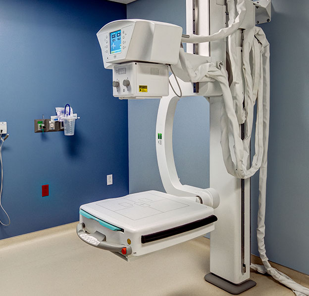

The technologist then moves the machine to aim the beam of radiation at the right area. Your body is put against a flat box or table that holds the x-ray film. You’ll be asked to sit, stand, or lie down, depending the body part to be x-rayed. You may be given a gown or drape to wear. You’ll need to remove jewelry or other objects that might interfere with the image. You will undress to expose the part of the body to be x-rayed. Usually x-rays are taken by an x-ray technologist. What is it like having x-rays(s)? Standard x-rays Your health care provider also might give you instructions.Īlways be sure to tell your health care provider whether you have allergies to iodine or have had problems with contrast materials in the past. The radiology center will give you instructions. You may be asked not to eat anything or to prepare in other ways before the test (see the next section). Preparation for a contrast study depends on the test.

Other than removing metal objects that might interfere with the picture, no special preparation is needed before having a standard x-ray. Veins throughout the body, most often in the leg Upper GI series, barium swallow, esophagography, small bowel follow through

Lower GI (gastrointestinal) series, barium enema (BE), double-contrast barium enema (DCBE), air-contrast barium enema (ACBE) For most of these tests, the images can be captured either on x-ray film or by a computer.Īngiography, angiogram, arteriography, arteriogramĪrteries throughout the body, including those in the brain, lungs, and kidneys It will look bright white on the x-ray and outline the body part. The contrast material may be given by mouth, as an enema, as an injection (put in a vein), or through a catheter (thin tube) put into various tissues of the body. During a contrast study, you get a contrast material that outlines, highlights, or fills in parts of the body so that they show up more clearly on an x-ray. Tumors are usually denser than the tissue around them, so they often show up as lighter shades of gray.Ĭontrast studies provide some information that standard x-rays cannot. Organs that are mostly air (such as the lungs) normally look black. Soft tissues block less radiation and show up in shades of gray. Tissues that block high amounts of radiation, such as bone, show up as white areas on a black background. After passing through the body, the beam hits a piece of film or a special detector. Dense tissues such as bones block most radiation, but soft tissues, like fat or muscle, block less. Tissues in the body absorb or block the radiation to varying degrees. How do x-rays work?Ī special tube inside the x-ray machine sends out a controlled beam of radiation. And angiograms may be used to diagnose non-cancerous blood vessel diseases. Still, angiography is sometimes used to show the blood vessels next to a cancer so surgery can be planned to limit blood loss. For instance, in the past, angiography was often used to help learn the stage or extent of cancer, but now CT and MRI scans are most often used to do this. See Table 1 for more examples.ĭue to advances in technology, many contrast studies are being replaced by other scans, such as CT or MRI scans. Another contrast study, an intravenous pyelogram (IVP), uses a special dye to look at the structure and function of the urinary system (ureters, bladder, and kidneys). For instance, a lower gastrointestinal (GI) series, often called a barium enema exam, takes x-ray pictures after the bowel is filled with barium sulfate. Special types of x-ray tests called contrast studies use iodine-based dyes or contrast materials, like barium, along with the x-rays to make the organs show up on the x-ray and get better pictures. To learn more about them, see Mammogram Basics. Mammograms (breast x-rays) are a form of radiographic tests. Still, x-rays are fast, easy to get, and cost less than other scans, so they might be used to get information quickly. They can show some organs and soft tissues, but MRI and CT scans often give better pictures of them. X-rays are very good at finding bone problems. Radiographs, most often called x-rays, produce shadow-like images of bones and certain organs and tissues. (For names of contrast studies, see Table 1.) What do x-rays show? Contrast studies may require more preparation ahead of time and may cause some discomfort and side effects, depending on what kind you are having. X-rays are typically fast, painless, and there’s no special preparation needed. X-rays and other radiographic tests (also known as radiographs, roentgenograms, and contrast studies) help doctors look for cancer in different parts of the body including bones, and organs like the stomach and kidneys.


 0 kommentar(er)
